Skip to content

From the American College of Rheumatology

Rheumatology for Primary Care
  • Symptoms
    • Fever
    • Joint Pain
    • Muscle Pain and/or Weakness
    • Rashes
    • Raynaud’s Phenomenon
  • Diseases
    • ANCA-Associated Vasculitis
    • Behcet’s Syndrome
    • Crystalline Arthropathies
    • Drug and Medication-Induced Rheumatic Diseases
    • Giant Cell Arteritis
    • Henoch-Schönlein Purpura
    • IgG4-Related Disease
    • Juvenile Dermatomyositis
    • Juvenile Idiopathic Arthritis
    • Kawasaki Disease
    • Mixed Connective Tissue Disease
    • Multisystem Inflammatory Syndrome in Children
    • Myositis
    • Polyarteritis Nodosa
    • Polymyalgia Rheumatica
    • Rheumatoid Arthritis
    • Sarcoidosis
    • Sjogren’s Disease
    • Spondyloarthritis
    • Systemic Lupus Erythematosus
    • Systemic Sclerosis
  • Case Studies
    • ANCA-Associated Vasculitis
    • Behcet’s Syndrome
    • Crystalline Arthropathies
    • Giant Cell Arteritis
    • Henoch-Schönlein Purpura
    • IgG4-Related Disease
    • Juvenile Dermatomyositis
    • Juvenile Inflammatory Arthritis
    • Kawasaki Disease
    • Mixed Connective Tissue Disease
    • Myositis
    • Pediatric Fevers
    • Polyarteritis Nodosa
    • Polymalgia Rheumatica
    • Raynaud’s Phenomenon
    • Rheumatoid Arthritis
    • Sarcoidosis
    • Sicca/Sjogren’s Disease
    • Spondyloarthritis
    • Systemic Lupus Erythematosus
    • Systemic Sclerosis
  • Labs
  • Meds
    • Commonly Used Rheumatology Meds
    • Treatment Considerations
    • Addressing Patient Fears About Rheumatology Medications
    • Medications by Diseases
  • Resources
    • Resources
    • Vaccination Considerations – Pediatrics
    • Vaccination Considerations – Adults
    • Glossary of Terms
    • ACR Clinical Guidelines
    • ACR Education Center
    • Rheumatology Provider Directory

Spondyloarthritis Case Studies

Home ยป Spondyloarthritis Case Studies
Spondyloarthritis Case Studies
  • Case 1

  • Case 2

  • Case 3

  • Case 1

A 33 yo M with no significant past medical history presents to the primary care clinic with low back pain.

Questions to Ask

  • Inflammatory back pain that is worse in the morning or with inactivity, and better with use or activity?
  • Duration of symptoms?
  • Other joint complaints?
  • Other organ systems involved (e.g., eyes, skin, bowels)?
  • Prior treatments?

Clinical Presentation & History

  • Initially developed low back pain and stiffness several years ago
  • Previously responded well to occasional ibuprofen, but now having breakthrough symptoms despite scheduled use 3x per day
  • Symptoms worst in morning, begin to improve somewhat after 2-3 hrs, but re-worsen with inactivity
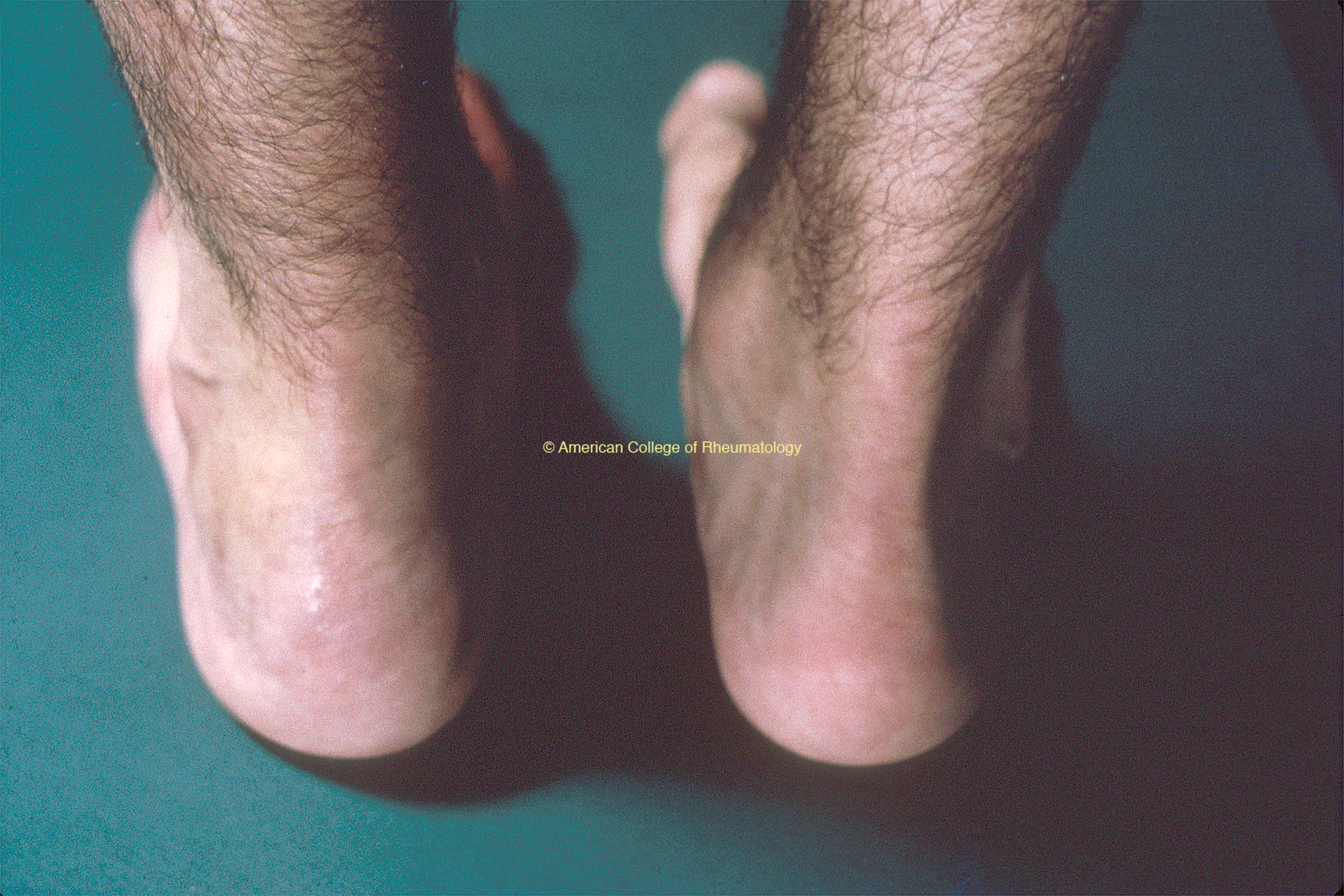
  • Also notes heel pain and swelling over the last few weeks (view image)

Diagnostic Workup

History suspicious for ankylosing spondylitis

Recommended Labs & Imaging
  • Labs:
    • CBC with diff, CMP, ESR, CRP
    • If high suspicion, consider HLA-B27 testing, hep B/C testing, and TB
  • Imaging: SI joint radiographs and heel X-ray
Test Results
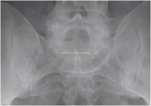
  • SI joint radiographs show bilateral sclerosis and erosions consistent with sacroiliitis (view image)
  • Labs show CRP 1.5x ULN and +HLA-B27
Diagnosis

Referral

Refer to rheumatology

Treatment & Further Workup

  • If no contraindications, start on full dose NSAID (e.g., naproxen 500 mg BID, meloxicam 15 mg daily, ibuprofen 800 mg TID)
    • If not improving after 2-4 wks, consider trial of alternate NSAID
  • Second line therapies include: TNF-inhibitor, IL-17 inhibitor, JAK inhibitor, etc.
  • Evaluate for other subspecialty needs:
    • Bowel disease
    • Uveitis
    • Psoriasis
  • Case 2

A 25 yo M with no significant past medical history presents to the primary care clinic with knee joint pain and swelling, rash, and dysuria + new penile discharge.

Questions to Ask

  • Inflammatory back pain that is worse in the morning or with inactivity, and better with use or activity?
  • Duration of symptoms?
  • Other joint complaints?
  • Other organ systems involved (e.g., eyes, skin, bowels)?
  • Prior treatments?

Clinical Presentation & History

  • New pain in both knees and ankles x2 weeks
    • Worse in the mornings
    • Improved with activity
  • Physical exam:
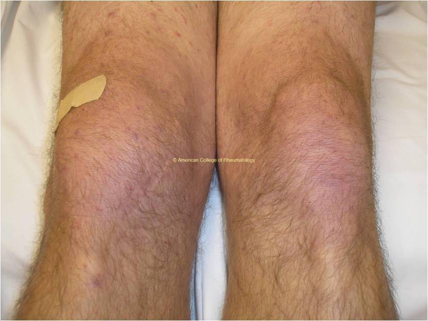
    • Right knee effusion (view image)
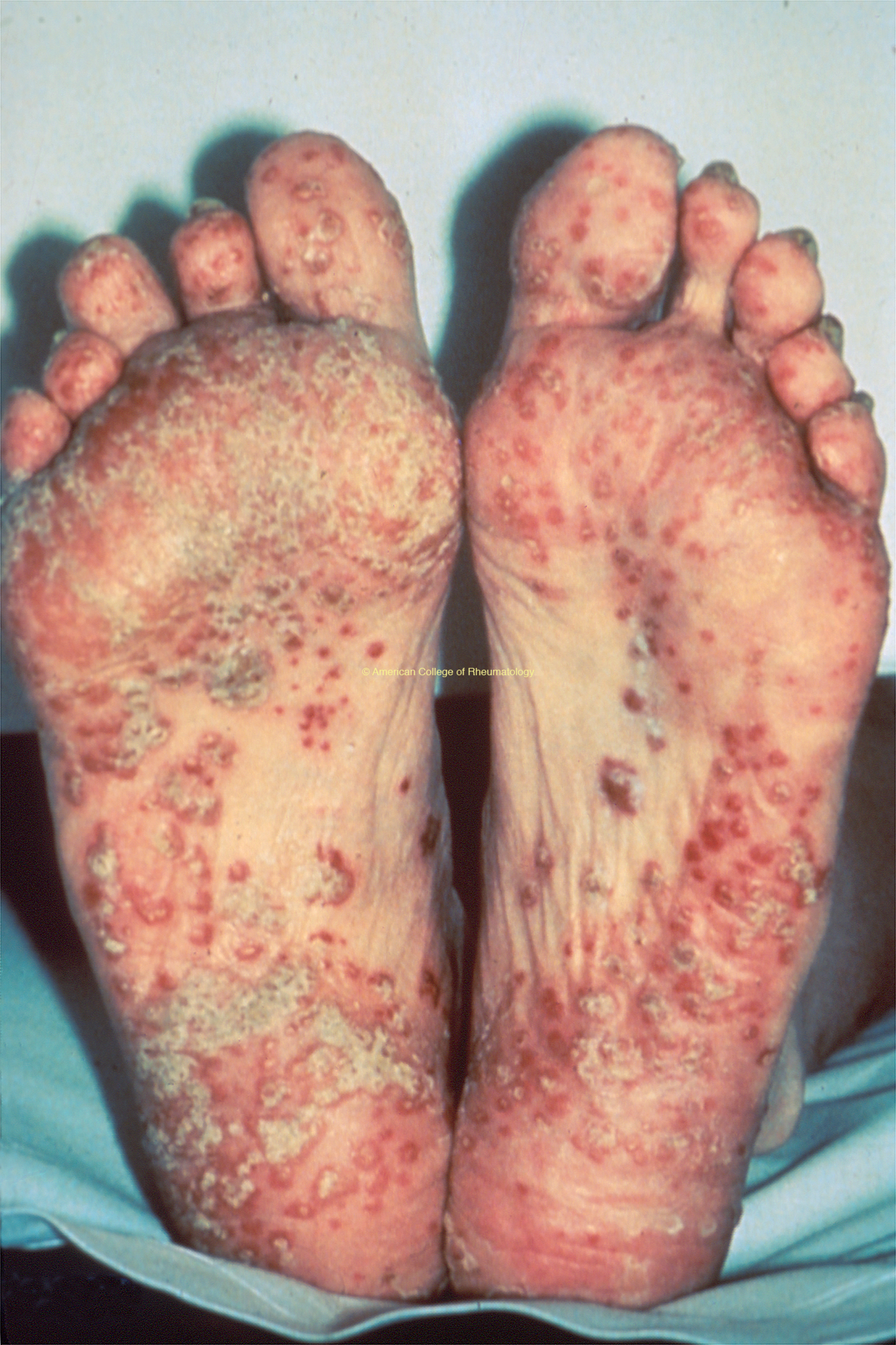
    • Hyperkeratotic rash on palms and soles (view image)
  • ROS
    • Dysuria
    • Penile discharge a few weeks ago after new sexual partner

Diagnostic Workup

Labs & Imaging
  • Labs: CBC with diff, CMP, ESR, urine chlamydia, gonorrhea, +/- other STIs
  • Imaging: plain radiographs of symptomatic joints
Test Results
  • Blood tests unremarkable
  • Knee x-ray:
    • Effusion
    • Soft tissue swelling
    • Otherwise normal
  • Urine PCR:
    • +Chlamydia
    • -Gonorrhea
  • Synovial fluid:
    • 18K WBCs/mm3 (58% PMNs)
    • Negative crystals/Gram stain/culture
Diagnosis

Treatment

  • Treat chlamydia infection
  • If no contraindications, start on full dose NSAID (e.g., naproxen 500 mg BID, meloxicam 15 mg daily, ibuprofen 800 mg TID)
  • Second-line treatments include steroids (systemic or intra-articular), sulfasalazine, etc.
  • Case 3

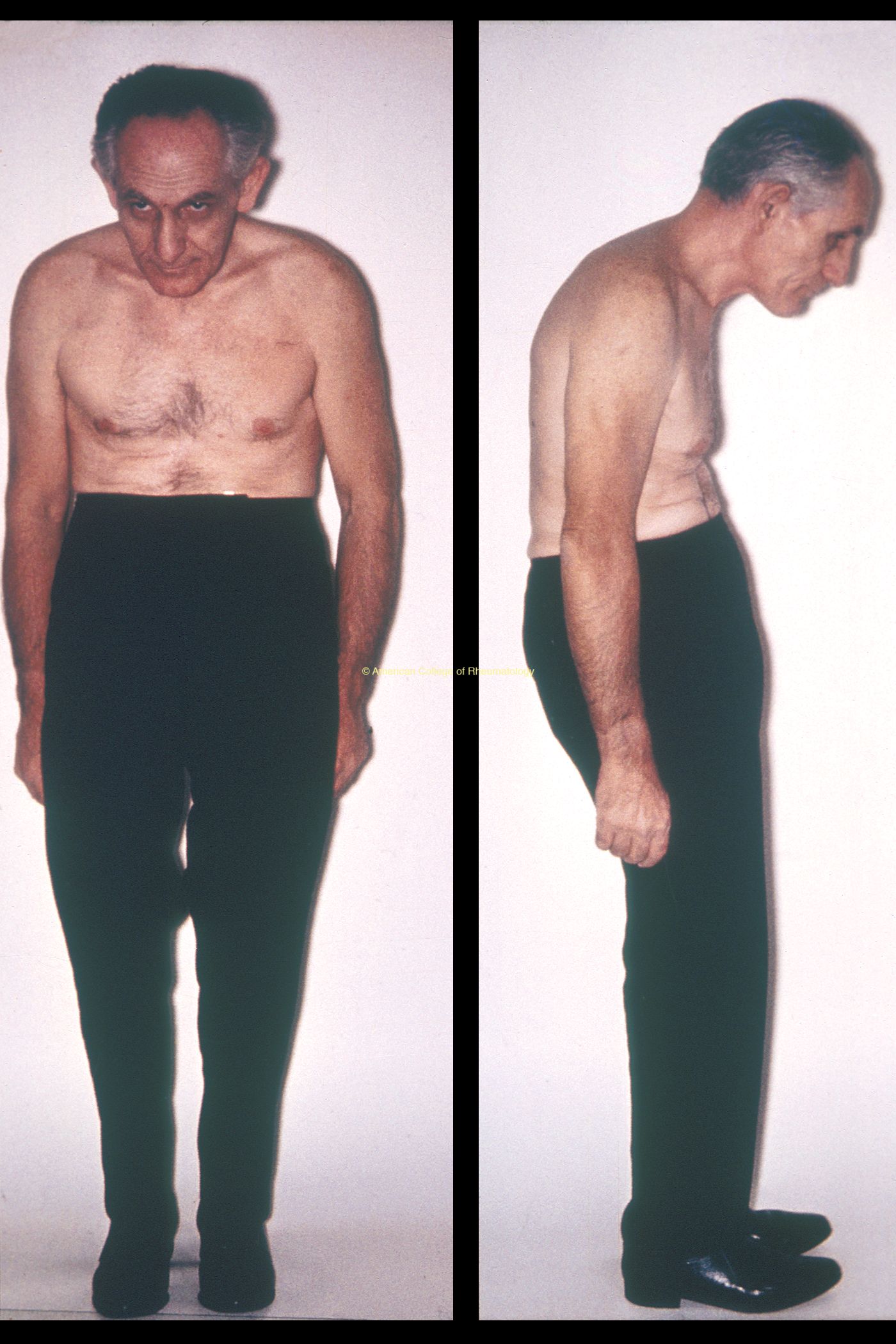
A 72 yo M with stooped posture (view image) presents to the clinic with breathing difficulties. He has not sought healthcare for many years.

Questions to Ask

  • Duration of symptoms?
  • Associated symptoms (e.g., cough, fever, orthopnea)?
  • Musculoskeletal complaints?

History & Physical Exam

  • ROS
    • Dyspnea and orthopnea worsening x several weeks
    • Swelling in both legs 
  • Physical exam
    • Rales at bilateral lung bases
    • Diastolic murmur
  • Chronic back pain starting in late 20s

Diagnostic Workup

Labs & Imaging
  • Basic bloodwork: CBC with diff, CMP
  • Dyspnea evaluation: EKG, CXR, TTE, etc.
  • Imaging: x-ray of his spine for stooped posture
Test Results
  • Evaluation shows acute decompensated heart failure, with TTE showing severe aortic regurgitation → valve surgery
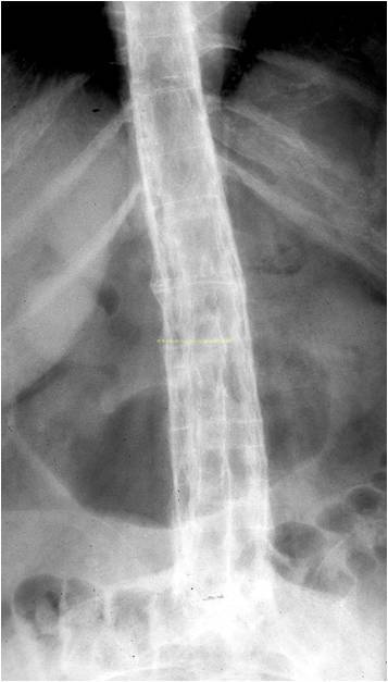
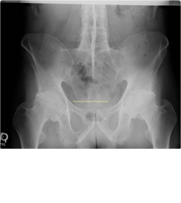
  • Spinal X-ray: fusion of his SI joints bilaterally (view image), as well as thin bridging syndesmophytes throughout his spine (view image)
Diagnosis

Referral

  • Refer to rheumatology

Management

  • Patient with known ankylosing spondylitis should be monitored for this complication and referred as necessary in order to prevent advanced disease and decompensation.

Clinical Pearl

  • Aortic regurgitation and aortic root disease are rare late complications of ankylosing spondylitis.

Diagnosis

Based on the diagnostic workup and clinical presentation, the patient has reactive arthritis.

Diagnosis

Based on the diagnostic workup and clinical presentation, the patient has spondyloarthritis.

Refer this patient to rheumatology.

Diagnosis

Based on the diagnostic workup and clinical presentation, the patient has ankylosing spondylitis.

Refer this patient to rheumatology.

Hyperkeratotic Rash on Soles of Feet

Numerous pustules are present on the feet of a patient with reactive arthritis. They begin as vesicles on erythematous bases and become sterile pustules. On the patient's right sole, the development of keratotic scales (keratoderma blennorrhagica) is most evident.

Numerous pustules are present on the feet of a patient with reactive arthritis. They begin as vesicles on erythematous bases and become sterile pustules. On the patient’s right sole, the development of keratotic scales (keratoderma blennorrhagica) is most evident.

Knee Swelling

Shows effusion in the right knee joint

Effusion in the right knee joint

Heel Pain & Swelling

Shows heel swelling

X-Ray Showing Sacrolitis

Radiograph showing bilateral erosive sacroiliitis with sclerosis of both the iliac and sacral surfaces. The iliac side is protected by a thin layer of fibrocartilage and is at increased risk for involvement in comparison to the sacral side, which is composed of hyaline cartilage.

Radiograph shows bilateral erosive sacroiliitis with sclerosis of both the iliac and sacral surfaces. The iliac side is protected by a thin layer of fibrocartilage and is at increased risk for involvement in comparison to the sacral side, which is composed of hyaline cartilage.

Stooped Posture

Left: Frontal view of a patient with ankylosing spondylitis (Marie-Strümpell disease) demonstrating the characteristic upward gaze of the eyes when looking straight ahead, necessitated by the flexion deformity of the neck. These postural changes are typical of the more advanced forms of this disease. Right: A lateral view of the same patient demonstrating forward protrusion of the head, flattening of the anterior chest wall, thoracic kyphosis, protrusion of the abdomen, and flattening of the lumbar lordotic curvature. Slight flexion of the hips is also present due to hip involvement. The partially fixed thoracic cage is primarily responsible for atrophy of the chest muscles.

Left: Frontal view of a patient with ankylosing spondylitis (Marie-Strümpell disease) demonstrating the characteristic upward gaze of the eyes when looking straight ahead, necessitated by the flexion deformity of the neck. These postural changes are typical of the more advanced forms of this disease. Right: A lateral view of the same patient demonstrating forward protrusion of the head, flattening of the anterior chest wall, thoracic kyphosis, protrusion of the abdomen, and flattening of the lumbar lordotic curvature. Slight flexion of the hips is also present due to hip involvement. The partially fixed thoracic cage is primarily responsible for atrophy of the chest muscles.

Spinal X-Ray

Lumbar and thoracic radiograph from a patient with ankylosing spondylitis showing classic ossification of the longitudinal ligaments symmetrically in a railroad track pattern leading to ankylosis of the vertebral bodies.

Lumbar and thoracic radiograph from a patient with ankylosing spondylitis showing classic ossification of the longitudinal ligaments symmetrically in a railroad track pattern leading to ankylosis of the vertebral bodies.

Pelvic X-Ray

Shows anteroposterior view of the pelvis of a 32-year-old male demonstrating advanced changes of ankylosing spondylitis. The sacroiliac joints are fused, there is diffuse symmetric narrowing of the hip joint spaces, and there are extensive syndesmophytes present in the lower lumbar spine.

This anteroposterior view of the pelvis of a 32 yo M demonstrates advanced changes of ankylosing spondylitis. The sacroiliac joints are fused, there is diffuse symmetric narrowing of the hip joint spaces, and there are extensive syndesmophytes present in the lower lumbar spine.

Related Links

Spondyloarthritis Overview


ACR Axial Spondyloarthritis Guideline

We Want Your Input

Find what you needed? We would love your feedback so that we can ensure the site meets the needs of primary care providers.

Give Feedback
Primary Care2024-05-21T14:01:43+00:00

American College of Rheumatology Information

About ACR

ACR Contacts

Authors

This product was funded by a medical grant from Pfizer. The content was written by a physician work group. See Authors

© Copyright American College of Rheumatology 2025 | All rights reserved | See ACR Policies

Page load link
Go to Top