About Myositis
Myositis is an inflammation of the muscles.
- Myositis abbreviations to know:
- Ages of onset:
-
Dermatomyositis > polymyositis can be associated with malignancy
Proximal muscle WEAKNESS >> pain
Clinical Phenotypes
- PM
- DM: myositis with rash
- CADM: typical skin rashes of DM, without weakness
- JDM: juvenile onset
- IBM:
- Proximal+distal mm
- Asymmetrical
- Inflammation and degeneration
- IMNM:
- Necrotizing myositis
- HMG-CoA reductase antibody in >90%, often with statin use
DM Rashes
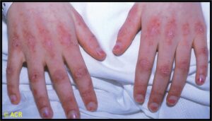
- Gottron’s papules (PIPs) (view image)
- Gottron’s sign
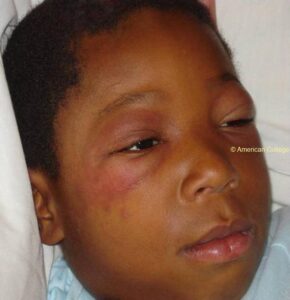
- Heliotrope rash (around eyes) (view image)
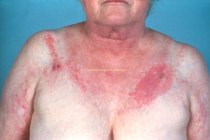
- Shawl sign (view image)
- Photosensitivity
Antisynthetase Syndrome
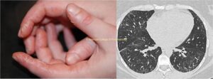
- Myositis + skin (mechanic hands) + ILD (view images)
- Arthritis
- Raynaud’s
Not everyone will have all of these manifestations.
Other Organs
- Pulm (interstitial lung)
- GI (dysphagia)
- Cardiac (myocarditis)
Lab Workup
- Creatine kinase high
- Aldolase high (some)
- ESR/CRP high
- AST/ALT elevation
- Myoglobinuria
- +ANA in a subset
- Myositis-specific antibodies such as Mi2, MDA5, NXP2, PM-SCL, SRP, TIF-1, anti-HMG CoA reductas per rheumatology
Imaging
- Muscle MRI can show muscle edema of affected muscles
- Chest CT if ILD is suspected
- ILD CT protocol = high resolution, without contrast, supine and prone views with deep inspiration and expiration
- Cardiac imaging (TTE, cardiac MRI, cardiac PET) to assess for cardiac muscle involvement if myocarditis is suspected
Additional Tests
- EMG shows myopathic changes
- High sensitivity, low specificity
- Muscle biopsy can be diagnostic
- Swallow study, if dysphagia is reported
- NIFs/FVC to assess respiratory musculature
- Further evaluation can be performed with MEP and MIP in full spirometry to differentiate extra thoracic from intrathoracic cause of restrictive lung involvement (i.e. ILD vs diaphragmatic involvement from myositis)
Rheumatic
- Polymyalgia rheumatica
- Overlap with systemic lupus erythematosus, mixed connective tissue disease, systemic sclerosis, etc.
Endocrinologic
- Diabetes mellitus
- Hypo-/hyper-thyroidism
- Adrenal insufficiency
Viral Infections
Neuromuscular
- Muscular dystrophies
- Neuromuscular junction disorder
- Denervating disorders
- Hereditary myopathies that can be adult onset (e.g., desferlinopathies)
Metabolic Myopathy
Rhabdomyolysis
Drug Toxicity
- Glucocorticoids
- Statins
- Colchicine
- Cocaine, alcohol
Electrolyte Imbalances
- Hyper/hypokalemia
- Hyper/hypocalcemia
Fibromyalgia
Initial Treatment
- Steroids: PO/IV prednisone equivalent to 1-2 mg/kg/day, then slow taper over months
- IVIG in select cases ((life threatening weakness, oropharyngeal dysphagia, respiratory involvement)
Long-Term Immunosuppression per Rheumatology
- Azathioprine
- Methotrexate
- Mycophenolate
- Cyclophosphamide
- Rituximab
- Tacrolimus
- JAK-inhibitors – dermatomyositis only
PT
Physical therapy and rehabilitation are very important to continue long term.
Monitoring
- Labs to follow:
- Muscle strength
- Monitor for signs of lung disease
- Follow up with PFTs + lung imaging
- Screen for underlying malignancy based on age-recommendations and risk stratification to the International Guideline for Idiopathic Inflammatory Myopathy-Associated Cancer Screening
Prognosis
- Symmetrical, proximal muscle weakness = hallmark of inflammatory myositis. If pain >>> weakness: consider alternate etiology (rhabdo, endocrinopathy)
- Classic rash distributions (Gottron’s papules over dorsum knuckles, heliotrope rash, etc.) with muscle weakness should prompt workup for dermatomyositis
- Vast majority of JDM cases have skin involvement
- HyperCKemia doesn’t equate to myositis:
- Different sexes and races can have different “normal limits”
- Exercise and dehydration can also impact CK levels
- Inclusion body myositis:
- Most common myositis of elderly
- Insidious weakness of hand flexors (FDP) and knee extensors (quadriceps); findings can be asymmetric initially
- Remember distal and proximal muscle weakness with neuropathic features
- Dermatomyositis and antibodies to NXP and TIF1 gamma in adults highly associated with malignancy, but dermatomyositis rarely associated with cancer in children


